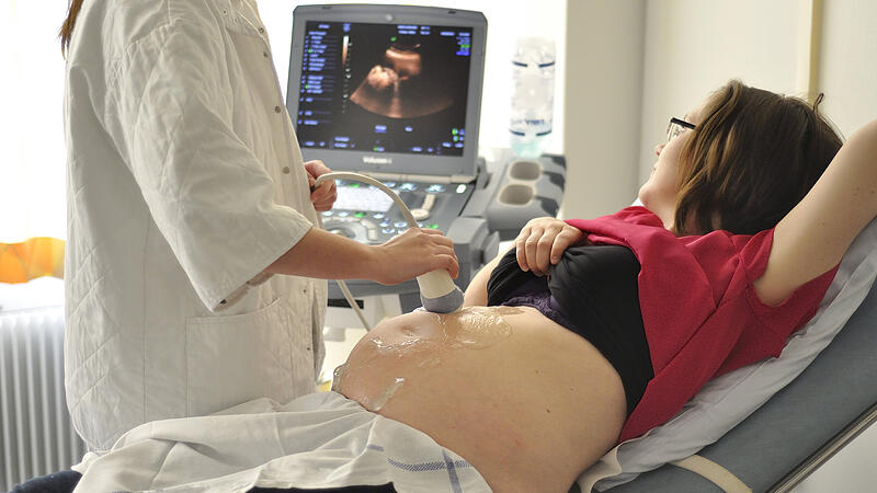
Prenatal ultrasound examination is an important method to assess the growth of a fetus. Malformations of the heart can also be diagnosed. The better the imaging, the more precise this is possible.
The research and production center of GE Healthcare is considered a specialist in women’s health. In order to further optimize the high-end ultrasound devices manufactured in Zipf, the company draws on the expertise of the researchers at the Center of Excellence for Medical Technology at the Upper Austrian University of Applied Sciences in Linz.
Very complex project
“We will use artificial materials to develop a simulator that reproduces the different layers of tissue in a womb, including the amniotic fluid, placenta and fetus, including the beating heart, as realistically as possible,” says Andreas Schrempf, who heads the research group. “We worked with GE Healthcare in a preliminary project and identified those materials that are similar to human tissue.” Now it’s time for implementation. “In fact, the fetal heart is the biggest challenge,” says Gerald Seifriedsberger, General Manager of GE Healthcare Austria. This is where doctors would recognize almost 80 percent of potential deformities in the early stages – a prerequisite for bringing about improvements with medication. At this point, the heart is one to three millimeters in size and is beating at 150 beats per minute. There are conventional ultrasound phantoms, but these are very standardized and not similar to human tissue.
Source: Nachrichten


