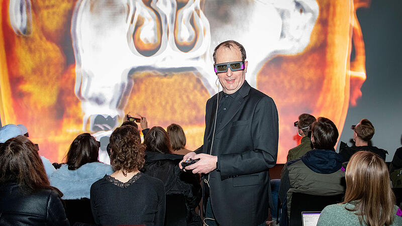
The Virtual Anatomy project of the Johannes Kepler University and the Ars Electronica Center was nominated in two categories for the E&T Innovation Award 2022 in London: “Best Emerging Technology of the Year” and “Most Innovative Solution in Digital Health and Social care”. The first took first place, the second took second place.
JKU Rector Meinhard Lukas accepted the award in London. The award shows “that the courage to tread innovative paths is worthwhile in every respect”. Lukas is “very proud that our joint project” continues to attract international attention after being nominated for the German Future Prize 2017. And Roland Haring, Technical Director of the Ars Electronica Futurelab, is also very happy: “We are delighted that our cooperation is not only revolutionizing medical teaching from Austria, but has now also been recognized internationally with the E&T Innovation Award.”
Immerse yourself in people
“Virtual Anatomy” has been used at the medical faculty of the Kepler University since last year. It combines MRI and CT data from real patients in a completely new way. Students and teachers can immerse themselves in the photorealistic images in 8K resolution. The teacher can rotate them freely using a controller and zoom into the smallest structures. The project was initiated by Franz Fellner, the dean of the medical faculty and head of the central radiology institute at the Kepler University Hospital. “With Virtual Anatomy we teach in a completely new way – modern, three-dimensional, calculated from CT and MR cross-section images,” he says.
In 2015, the 16 by 9 meter Deep Space 8K in the Ars Electronica Center was turned into a lecture hall for the first time with “Cinematic Rendering” technology from the Siemens Healthineers company. This was followed by regular lectures for medical students as well as presentations on anatomy for laypeople and live broadcasts on operations.
“Door to the Future”
Horst Hörtner, Managing Director of the Ars Electronica Futurelab, is certain that this will open the “door to the future of university education”. Since the images shown come from real patients, the natural-looking visualization makes it easier for doctors and their patients to explain damage to the body, the diagnosis of a disease or the course of a planned operation. Of course, the same is also useful for medical students, therapists or nursing staff, because the Virtual Anatomy allows previously unthinkable insights into anatomical details. For example, it depicts fibers in the human brain with a level of detail that would otherwise not be possible.
“I am extremely pleased that this project has now received an award,” says Fellner. This confirms “our way of combining digital and analogue teaching in a meaningful way.”
Source: Nachrichten


